Closely intertwined

Breast cancer and cardiovascular risk

Review Article
Issue
2023/03
DOI:
https://doi.org/10.4414/cvm.2023.02275
Cardiovasc Med. 2023;26(03):100-106

Affiliations
a University Heart Centre, University Hospital of Zurich, Zurich, Switzerland;Departments of b Medical Oncology, c Cardiology, and d Biomedicine, University Hospital Basel, Basel, Switzerland; e University of Basel, Basel, Switzerland


Published on 17.05.2023

Summary

With the continuous improvement of therapies against breast cancer, the long-term onset of cardiovascular disease (CVD) is becoming increasingly relevant for both cardiologists and oncologists. Not only CVD arises from known cardiac side effects of several anti-cancer therapies, but cancer itself seems to promote CVD. On the other hand, there is increasing evidence that CVDs such as myocardial infarction and heart failure predispose to future development of cancer. The fast-developing field of cardio-oncology aims to characterise cancer patients in order to implement effective tools for surveillance and prevention of cardiovascular adverse events and to raise awareness for the increased cancer incidence in patients with CVD. The aim of this review is to highlight cardiovascular side effects and toxicities of some of the most important breast cancer therapies and to provide an overview of what is known on the complex interplay between CVD and breast cancer.
Keywords: Cardio-oncology, breast cancer, cardiotoxicity, cardiovascular disease, heart failure, risk factors

Introduction

Due to impressive advances in cancer therapies, survival of women presenting with early breast cancer continuously improved over the past decades. The reduction in cancer-related mortality has led to a corresponding increase in cardiovascular diseases (CVD), which currently represent the leading cause of morbidity and mortality among breast cancer survivors in Western countries [1]. Besides cardiovascular side effects of anti-cancer therapies, the increase in CVD is both influenced by ageing and shared risk factors between CVD and breast cancer, such as obesity and smoking [2].
Cardiovascular side effects of breast cancer therapies are diverse and include, among others, an increased risk of heart failure associated with anthracyclines and human epidermal growth factor-receptor 2 (HER2) inhibitors, the promotion of atherogenesis associated with long-term oestrogen suppression and of coronary artery disease due to (particularly left-sided) chest radiation [2-4]. In addition, there is increasing evidence that CVDs such as myocardial infarction might directly promote the development of breast cancer due to complex cross-disease mechanisms involving a deranged immune answer [5].
Cardio-oncology represents a new developing branch of cardiology, which aims to make cancer treatments safer from a cardiovascular perspective. A better understanding of the pathophysiological mechanisms behind CVD and cancer will allow not only the development of more efficient tools for surveillance and prevention of cardiovascular adverse events, but also raise the awareness that patients affected by CVD might in the future present with cancer.
The aim of this review is to highlight the cardiovascular-related adverse effects of some of the most important breast cancer therapies and to provide an overview of the common pathways influencing the complex interplay between CVD and breast cancer. The first section summarises the epidemiology and the proposed pathophysiological mechanisms behind cardiotoxicity of drugs used in breast cancer treatment, including anthracyclines, HER2-therapies, radiotherapy and hormone therapy. In the second part, correlation of cardiovascular risks and breast cancer related outcomes are discussed, with a focus on the shared risk factors between CVD and breast cancer. Finally, evidence of how CVD may influence and predispose for the development of cancer is presented.

Cardiotoxicity of breast cancer therapies

Anthracyclines

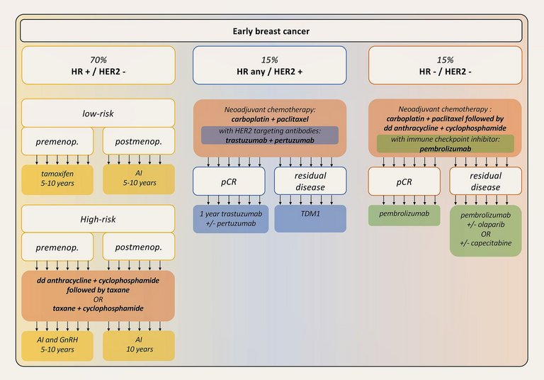
Anthracyclines are highly effective in the treatment of various solid tumours and, as such, still represent the cornerstone of mostly neoadjuvant chemotherapeutic regimens for high-risk early breast cancer (fig. 1) [6]. Although highly effective, their use has been limited due to irreversible, dose-dependent cardiac damage, which may negatively affect the prognosis despite effective cancer treatment. Therefore, appropriate risk stratification and cardiovascular monitoring before starting as well as during anthracycline therapy are recommended to guarantee optimal clinical outcome [7].
Figure 1: Overview of the different types of breast cancer and their therapies. Simplified breast cancer-related risk stratification and related, most used therapies. AI: Aromatase inhibitor; dd: dose-dense; GnRH: Gonadotropin-releasing hormone agonists; HER2: Human epidermal growth factor receptor 2; HR: Hormone receptor; pCR: Pathological complete remission; TDM1: Trastuzumab emtansine.
Understanding the pathophysiological mechanisms behind anthracycline-induced cardiotoxicity is critical to develop appropriate prevention and treatment strategies. Initially, anthracyclines were believed to cause left ventricular dysfunction mainly by directly promoting oxidative stress via iron-dependent generation of reactive oxygen species (ROS) [8]. Later on, further data from animal models revealed that toxicity-related mechanisms depend on the effects of doxorubicin on the type II topoisomerase [9]. Type II topoisomerase plays an important role in DNA replication by causing transient breaks and reunion of double-stranded DNA. Through binding to type II topoisomerase, doxorubicin inhibits its activity, which leads to activation of a DNA damage response and apoptosis in fast replicating cells. However, doxorubicin-bound type II topoisomerase also suppresses the transcription of genes involved in oxidative phosphorylation and electron transport, thus directly interfering with mitochondrial biogenesis and function resulting in increased ROS generation and altered mitochondrial biogenesis in cardiomyocytes [9]. Since cardiac involvement is mainly related to the interaction between doxorubicin and type II topoisomerase, patients with higher expression of this isoform in cardiomyocytes are more likely to develop cardiotoxicity. In humans, anthracyclines have a dose-dependent toxicity. In early stages, anthracyclines may lead to reversible myocyte damage and dysfunction, mainly related to oxidative stress, so that an improvement of the left ventricular ejection fraction (LVEF) is expected once the therapy is withdrawn and heart failure therapy is started. Once myocyte death occurs, the damage becomes irreversible, leading to a dose-dependent reduction in left ventricular (LV) mass likely caused by apoptosis and myocyte atrophy [10, 11]. A reduction in the LV mass index represents an adverse prognostic factor for cardiovascular outcomes [10, 11].
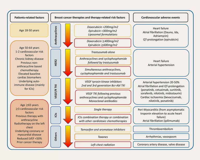
The cumulative life-time dose of anthracyclines is conventionally limited to ≤550 mg/m2 doxorubicin (or the calculated equivalent in case other anthracyclines are used) based on epidemiological data showing a dose-corresponding incidence of heart failure of 7% at this threshold [2]. The diagnosis of anthracycline-related cardiotoxicity relies on imaging (typically measurement of LVEF by transthoracic echocardiography and LV global longitudinal strain). Based on increased imaging sensitivity, the incidence of heart failure for the same cumulative dose is 26%, resulting in a reduction of the recommended cumulative lifetime dose to 400-450 mg/m2 (fig. 2) [12]. Although such doses are usually not reached in breast cancer patients, lower doses may cause cardiotoxicity in vulnerable patients and combination with other potentially cardiotoxic therapies, namely HER2-inhibiting drugs or chest radiation may also increase the risk of cardiovascular toxicity. Elevated troponin values before anthracycline therapy may help identify those patients, who will be at highest risk to experience late-onset cardiotoxicity [2].
Figure 2: Cardiotoxicity of the most important therapies against breast cancer. ICIs: Immune checkpoint inhibitors; LVEF: Left ventricular ejection fraction; TKI: Tyrosine kinase inhibitors; VEGF/R: Vascular endothelial growth factor/receptor.
Several prevention and screenings strategies have been proposed to reduce the burden of anthracycline-related cardiotoxicity while at the same time guaranteeing effective cancer treatment. Mostly, proposed strategies include administration of anthracyclines via continuous infusion, as liposome-encapsulated drugs or switching to less cardiotoxic anthracyclines such as epirubicin or idarubicin [13]. Furthermore, simultaneous use of cardioprotective drugs such as dexrazoxane, a strong topoisomerase inhibitor itself, which prevents anthracyclines from binding to the type II topoisomerase-complex, may be employed [13]. This therapy is however costly, only approved in adults with progressive solid tumours who already received >300 mg/m2 doxorubicin and might reduce anthracycline’s anti-cancer efficacy [13]. Although anthracycline-related cardiotoxicity was initially defined as irreversible, myocardial recovery after anthracycline-induced injury may occur following early therapy with enalapril and carvedilol [14]. However, studies were affected by short-term follow-ups and it remains unclear whether the positive results derive from primary cardioprotection or rather from variation in haemodynamics [13].
Screening strategies are the cornerstone to monitor anthracycline therapy. Transthoracic echocardiography with strain analyses for detection of ventricular damage prior to manifest LV dysfunction has emerged as a promising method. With this respect, cardiotoxicity is defined as the onset of symptomatic cardiac dysfunction with heart failure symptoms independently from LVEF during the course of anthracycline therapy or an asymptomatic cardiac dysfunction defined as new LVEF reduction <50% or a new relative reduction in global longitudinal strain of >15% from baseline and/or a new rise in cardiac biomarkers (troponin or natriuretic peptide) despite a LVEF ≤50% [15, 16]. Cardiac biomarkers of myocyte damage such as troponin are helpful, but their specificity is limited and the timing as well as the exact cut-off to define anthracycline-related cardiotoxicity have not been defined in large prospective trials yet [17]. However, according to the new European Society of Cardiology (ESC) guidelines, measurement before each cycle of anthracyclines is recommended [15]. Further biomarkers such as NT-proBNP may identify patients at higher risk of developing cardiotoxicity and may be more helpful for long-term surveillance [17].

Cyclophosphamide

Cyclophosphamide is an alkylating agent with cytostatic properties, which is related to the onset of cardiomyopathy when administered in high doses [18]. Cyclophosphamide is metabolised via cytochrome P450 to aldophosphamide, which decomposes to phosphoramide mustard and acrolein. The first is an active neoplastic agent which induces apoptosis by interacting with DNA, whereas the latter is a toxic metabolite which causes toxicity in the myocardium, cardio-fibroblasts and endothelial cells [19]. At a lower dose, cyclophosphamide results mainly in a therapeutic immunosuppression, whereas the cardiotoxic effect is particularly evident at an antineoplastic therapeutic dose of 120-200 mg/kg [20]. Cyclophosphamide is most commonly administered in combination with an anthracycline (doxorubicin or epirubicin) in neoadjuvant treatment of high-risk early breast cancer (fig. 1) [21, 22]. Prior to recent de-escalation studies, standard treatment for all women with breast cancer included both therapies. Presently, for lower risk patients, anthracyclines are omitted and cyclophosphamide is combined with docetaxel. However, high-dose cyclophosphamide is not used as breast cancer therapy. Therefore, no routine surveillance of breast cancer patients receiving cyclophosphamide is needed.

Taxanes

Taxanes are microtubule-stabilising drugs, which act on the cellular mitotic spindles and induce cell death during mitosis [23]. In current practice, taxanes are used ubiquitously in all chemotherapy regimens for early breast cancer (fig. 1). In triple negative and HER2-positive breast cancer docetaxel three-weekly or paclitaxel weekly are combined with carboplatin. In hormone receptor positive HER2-negative breast cancer combination of taxane and cyclophosphamide is standard of care [24]. Due to the complexity of the therapeutic regimens and multidrug interactions, it is often difficult to define the exact role of taxanes alone when defining cardiotoxicity. Indeed, taxanes relevantly interact with anthracyclines and increase their plasma levels, promote the formation of more cardiotoxic metabolites and enhance the global cardiotoxicity [25, 26]. Thus, particular caution is warranted when both therapies are administered together and the administration of anthracycline in separate infusions before taxanes, particularly paclitaxel, is recommended [2]. Taxanes are, however, known for their proarrhythmogenic effect as well as for inducing vasospasms in the coronary arteries (fig. 2) [15].

HER2 inhibitors

Inhibitors of HER2 such as antibodies (trastuzumab, pertuzumab), antibody-drug conjugates (trastuzumab emtansine, trastuzumab deruxtecan) or small molecule tyrosine kinase inhibitors (lapatinib, neratinib, tucatinib) are used either as monotherapy or in conjunction with chemotherapies for the treatment of HER2-positive breast cancer (fig. 1). Contrary to anthracyclines, trastuzumab-related cardiotoxicity mainly develops during treatment, is mostly reversible, not cumulative dose-related, albeit increasing with long-time use, and long-term follow-up data (up to ten years after treatment) are reassuring [2, 27]. If trastuzumab was administered right after anthracycline containing regimens, a summative effect with respect to the incidence of heart failure has been observed (fig. 2) [2]. Antibodies such as trastuzumab bind to the extracellular domain of HER2 inhibiting its activity, whereas tyrosine kinase inhibitors enter the cell and target the HER2 intracellular kinase domain [28]. The aetiology behind trastuzumab-induced cardiotoxicity is mainly related to myocyte dysfunction in the absence of irreversible injury such as major cell death as shown in biopsy studies [29]. HER2 inhibitors block HER2 receptors on the surface of cardiomyocytes interfering with signalling pathways responsible for cell repair in response to cellular stress as well as with antiapoptotic pathways [29]. These effects explain the enhanced cardiotoxicity of the combination of chemotherapies with anthracyclines and HER2 inhibitors.
Since cardiotoxicity related to adjuvant HER2-blockade mainly occurs during treatment, several surveillance protocols have been developed [30]. Heart failure is the main reported side effect with high incidence rates up to 27% when administered in combination with anthracyclines in women with metastatic breast cancer, but as low as 3% when given alone [31, 32]. Older age, above 50 years, a lower baseline LVEF ≤55% or presence of traditional cardiovascular risk factors are associated with an increased risk [33]. The role of troponins in the surveillance during HER2 directed therapy is questioned, since myocardial necrosis is rare and not directly caused by these drugs [17]. As no population-based studies investigating the long-term risk of cardiac dysfunction in women treated with HER2-inhibitors alone are available, there are no clear protocols for risk stratification and recommendations about preventive strategies in these patients [34]. However, a recent meta-analysis showed a beneficial effect of beta blockers, angiotensin converting enzyme inhibitors and angiotensin receptor blockers on LVEF under trastuzumab and, to a lesser extent, also anthracycline therapy [35]. In general, repeated echocardiographic and biomarker assessment for early detection of cardiotoxicity is recommended during and after treatment according to risk category and should be continued long-term, as HER2-related toxicity may increase over time [15, 30].
Recently, novel antibody-drug conjugates (ADC) such as trastuzumab emtansine (TDM1) and trastuzumab deruxtecan (T-DXd) entered the clinical stage. Highly potent but very toxic cytostatic agents are coupled to an antibody targeting HER2 facilitating an increase of intratumoural drug levels and a decrease of off-target toxicity. T-DXd is used as second line therapy in HER2 positive and selected HER2 “low” expressing metastatic breast cancer [36, 37]. In early HER2 positive breast cancer, adjuvant treatment escalation to TDM1 is standard of care if a complete pathologic response was not achieved after neoadjuvant therapy (fig. 1) [38]. Whereas clinical experience with these kinds of drugs is still limited and only little is known about their potential cardiotoxicity, results on tumour efficacy are encouraging [37, 39].

Radiation

Radiation therapy is associated with a higher long-term risk for cardiovascular adverse events and is particularly increased in case of left-sided breast radiotherapy (fig. 2) [40]. Left-sided radiotherapy might affect cardiac structures and function in several ways, from pericardial and myocardial fibrosis resulting in constriction and restriction to alteration of the valvular apparatus, a more rapid progression of coronary artery disease, up to the involvement of the heart conduction system [17]. The scope of the cardiac involvement depends on the radiation dose, the involved heart region as well as on pre-existing comorbidities and concomitant cardiotoxic therapies [2]. In particular, radiation therapy induces direct endothelial damage, which favours the onset of atherosclerotic coronary plaque formation and is related to a general increase of death from ischaemic heart disease [41, 42]. Furthermore, radiation may induce fibrosis, calcification and dysfunction of the heart valves [43].
Over the past years, several strategies were developed to protect the heart, such as lowering the total radiation dose, using more precise radiation fields, the deep inspiration breath hold technique and shielding of the myocardium [34]. The application of these preventive techniques has led to a substantial decrease in the risk of cardiovascular death in women who underwent radiotherapy for breast cancer [42].

Hormone therapy

Endocrine hormonal therapy is the cornerstone of adjuvant cancer therapy in breast cancer expressing hormone-sensitive receptors (such as oestrogen or progesterone receptors, fig. 1) [44]. Hormonal therapies interfere with oestrogen-stimulated growth of normal and cancerous cells. Depending on their exact molecular target, they exhibit several cardiovascular side effects. In adjuvant settings, hormone therapies might be administered for more than five years, thus emphasising the importance of the evaluation of their cardiovascular side effect profile.
Tamoxifen represents the first-line endocrine intervention developed for early-stage hormone receptor-positive breast cancer in premenopausal women, in whom it efficiently interferes with oestrogen signalling by binding to oestrogen-receptors [45]. Although tamoxifen has lipid-lowering effects with reduction in total serum and low-density lipoprotein cholesterol, long-term clinical trials failed to demonstrate a protective effect with respect to cardiovascular outcomes [46]. On the contrary, tamoxifen has been linked to higher rates of arterial thrombotic events (fig. 2) [44].
Aromatase inhibitors (AIs) represent the principal hormonal therapy for postmenopausal women alone or in combination with gonadotropin releasing hormone (GnRH) analogues in premenopausal women with high-risk features (fig. 1) [45, 47, 48]. AIs inhibit the endogenous oestrogen production by acting on the aromatase enzyme, which normally converts androstenedione to oestradiol. Adjuvant therapy with AIs is efficient in both postmenopausal women, where oestrogen production occurs mainly thorough aromatase in peripheral tissues, as well as in premenopausal women [49, 50]. Several trials compared cardiovascular outcomes in patients under AIs to patients treated with tamoxifen and found AIs to be associated with an increased cardiovascular risk [51, 52]. In contrast, in a randomised trial of AIs versus placebo after five years of tamoxifen, no increased cardiovascular risk was found [53]. Still, AIs have been associated with a higher rate of arterial thrombosis and stroke as well as some of them with an increase in blood lipids, despite a relatively low cumulative rate of adverse events [54]. However, the quality of these trials is often limited by lack of sufficient information about cardiovascular therapies and cardiovascular-related comorbidities, which might have influenced the results. Current guidelines recommend a baseline and annual assessment of cardiovascular risk based on the SCORE2 or SCORE2-OP and secondary prevention according to the degree of dyslipidaemia and/or arterial hypertension [15].
Another promising therapy are cyclin-dependent kinase 4/6 (CDK4/6) inhibitors, which target the growth of cancer cells and are currently used to treat hormone receptor-positive HER2-negative metastatic breast cancer in association with hormone therapies [55]. Whereas QT-time prolongation has been described for ribociclib, data on long-term cardiotoxicity is not yet available [56].

Immune checkpoint inhibitors

Immune checkpoint inhibitors (ICIs) consist of monoclonal antibodies, which block physiological immune brakes or “checkpoints” such as the cytotoxic T-lymphocyte-associated protein 4 (CTLA-4: Ipilimumab, tremelimumab), programmed cell death protein 1 (PD-1: Nivolumab, cemiplimab, pembrolizumab) or its ligand (PD-L1: Atezolizumab, avelumab, durvalamab) to activate T-lymphocytes that target the tumour [15]. Triple-negative breast cancer cells overexpress PD-L1, which renders them susceptible to ICI therapies [57]. Hence, addition of pembrolizumab to chemotherapy has recently been approved for treatment of early and metastatic PD-L1 positive triple negative breast cancer (fig. 1) [58, 59].
A potentially lethal side-effect of these drugs is related to the activation of T-cells that may also target other, non-cancerous tissues including the myocardium [60]. ICI-related myocarditis most frequently develops within the first twelve weeks of treatment and manifests with a wide range of signs and symptoms, ranging from asymptomatic troponin elevation to fulminant myocarditis with severe myocardial dysfunction [60, 61], though the diagnosis can be challenging in milder cases (fig. 2). Endomyocardial biopsy should be undertaken in case of uncertainty, as a positive diagnosis has relevant consequences due to the necessary discontinuation of a potentially curative therapy [15]. Multidisciplinary team discussion is warranted to assess the risks versus benefits of resuming therapy in cases of mild myocarditis [15]. Treatment consists of high-dose corticosteroids and, in case of a non-response, other immunosuppressive agents. Besides myocarditis, ICI can also lead to vasculitis (and therefore acute coronary syndrome) or pericarditis and rarely, Takotsubo cardiomyopathy may occur.

Cardiovascular risk and outcome of breast cancer patients

In the last decades, steady developments in cancer therapies for breast cancer have dramatically improved survival increasing deaths from other causes, including cardiovascular and cerebrovascular diseases. The diagnosis of cancer holds per se a worse prognosis in terms of cardiovascular mortality and morbidity [62]. In addition, according to an English retrospective cohort study, cardiovascular mortality “overruns” cancer mortality in >60 year-old survivors of nine different cancer types, including breast cancer [63]. According to several studies, breast cancer survivors have a 1.8-fold higher risk of cardiovascular death compared to cancer-free women in the long-term, irrespective of tumour stage, hormone receptor status or age at diagnosis and the risk is even higher in those receiving chemotherapy [64, 65]. Although older patients had the highest risk, suggesting a summative effect with traditional risk factors, even breast cancer with positive oestrogen-receptor expression was linked to an increased mortality [64]. In the Women’s Health Initiative study including older breast cancer survivors a higher incidence of heart failure with preserved EF (HFpEF) as compared to heart failure with reduced EF (HFrEF) has been observed. HFpEF was related to a twelve-fold higher risk for cardiovascular mortality [66]. On the other hand, the onset of HF was related to a non-significant two-fold higher risk for breast cancer mortality [66].
The pathophysiological mechanisms underlying this association are not well understood. In the Pathways Heart Study, an ongoing prospective cohort study, 13,642 women firstly diagnosed with invasive breast cancer were compared to 68,202 controls matched for age and ethnicity with no history of breast cancer in a 5:1 fashion [67]. Patients who received combined anthracyclines and trastuzumab had the highest risk of long-term cardiovascular adverse events defined as heart failure incidence and/or cardiomyopathy as compared to both controls and patients who were treated with monotherapy [67]. Interestingly, the risk proportionally increased from the lowest elevated risk in women treated with anthracyclines without trastuzumab, followed by those on trastuzumab without anthracyclines and, in line with other cohort studies, by those with combined therapy [67, 68]. Considering patients with previous traditional cardiovascular risk factors, the risk of rate of death from CVD and breast cancer is similar within the first five years after cancer diagnosis and, thereafter, death due to CVD dramatically increases reaching the highest prevalence in women 66 years or older [69]. Furthermore, anthracycline exposure has been related to a 1.8- up to 3-fold higher risk for arrhythmia, although this might itself represent a phenotype of an underlying cardiomyopathy [67, 69]. Accordingly, the recent SEER Medicare Analysis showed a higher incidence of atrial fibrillation in women with breast cancer as compared to women without cancer. This was associated with an increased cardiovascular mortality after one year follow-up [70]. Still, the increasingly investigated cancer therapy-related cardiotoxicity does not account alone for the enhanced cardiovascular mortality reported in large prospective epidemiological studies [67, 71].

Shared risk factors

Although large-scale population-based studies investigating the risk of CVD in breast cancer survivors have been inconsistent, a recent cohort study based on medical records found that long-term breast cancer survivors had a higher risk of CVD at 10-15 years from initial diagnosis [71]. Interestingly, the risk came to the same as for the general population after >15 years from diagnosis and, in general, an association between chemotherapy, hormonal therapy or radiotherapy with late-onset CVD was not observed [71]. Main predictors for CVD in this study were older age, obesity, a family history of CVD, a family history of breast cancer, low education and comorbidities according to the Charlson Comorbidity Index [71].
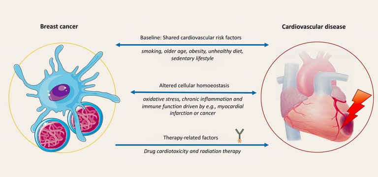
Indeed, breast cancer and CVD share many common risk factors such as smoking, older age, obesity and unhealthy diet rich in red or processed meats and high-fat dairy products (fig. 3) [28]. Different types of fat seem to differently affect the risk of breast cancer. In particular, a diet rich in n-3 polyunsaturated fatty acids has been associated with a decrease in both adverse cardiovascular events and, according to a meta-analysis, with a risk reduction of breast cancer [72]. However, another study indicates that this association may be primarily driven by a lower carbohydrate intake [73]. Alcohol consumption represents an important exception: although a light to moderate alcohol consumption has been associated with a risk reduction for CVD in a J-shaped fashion [74], it is associated with a continuous dose-dependent increase in the risk for particularly hormone receptor-positive breast cancer [75]. All in all, there is conflicting evidence on the link between dietary-related risk factors and the onset of CVD and breast cancer, as these associations are mainly based upon epidemiological studies, which fail to provide causation and are largely influenced by population-related cofounders [28].
Figure 3: Shared risk factors and disease interactions between breast cancer and cardiovascular disease. The Figure was partly generated using Servier Medical Art, provided by Servier, licensed under a Creative Commons Attribution 3.0 unported license.
© Servier Medical Art / Creative Commons
Similarly, a sedentary lifestyle has been associated with both an increased risk of CVD and breast cancer, whereas an active lifestyle has been associated with a lifetime risk reduction of breast cancer of 9-18%, with lower values in women who used hormone replacement therapy [76-78]. As in CVD, physical activity has a positive prognostic effect also in breast cancer survivors [79]. The pathophysiology behind this association is likely related to the effects of endogenous sex steroids and to the positive effects of physical activity on insulin sensitivity, oxidative stress, chronic inflammation and immune function [80].
Obesity, which is often strictly related to a sedentary lifestyle, is a known risk factor for CVD in a linear progressive fashion [76]. In contrast, although each 5 kg weight increase is associated with an 11% risk increase of breast cancer, this was only true for postmenopausal women, whereas no relationship was found for premenopausal women, in whom long-term obesity seems to have rather a protective effect [81]. On the other hand, short-term weight gain in the perimenopause has been associated with higher breast cancer risk, even after controlling for Body Mass Index [82].
Smoking is a well-established risk factor for CVD, however, epidemiological evidence about its role in breast cancer is inconsistent, likely due to heterogeneity in clinical studies and poor prospective studies [76, 83, 84]. Recently, a positive association among women who started smoking early in their adolescence has been reported [84]. In this study, Jones et al. observed a summative effect of smoking in the presence of a positive family history of breast cancer, likely linked to genetic variants in enzymes involved in metabolism of carcinogens such as N-acetyltransferase 2 [85].

When CVD promotes breast cancer

Cancer onset is favoured by adverse variation in cellular homoeostasis. However, the direct impact of CVD on breast cancer has been poorly investigated. Lately, new evidence from animal models has shed light on the influence of acute cardiovascular events such as a myocardial infarction on the development or progression of cancer. Myocardial infarction induces a local and systemic inflammatory host response by provoking myocyte necrosis [86]. After a myocardial infarction, β3-adrenergic signalling leads to the release of leukocytes and myeloid progenitor cells from the bone marrow, which regulate the local inflammatory response in the atheromatic ruptured plaque [86]. The locally activated proinflammatory mononuclear phagocytes release interleukin-1. This cytokine epigenetically reprograms myeloid cells in the bone marrow and in extramedullary sites toward an immunosuppressive state and induces systemic monocytosis. This event finally leads to deleterious cross-disease communication, enabling tumour growth (fig. 3) [86]. In fact, monocytes are among the key regulators of the tumour microenvironment and are responsible for the tumour’s immune evasion, neoangiogenesis as well as cell migration and proliferation [87]. The presence of these synergies between myocardial infarction and breast cancer was lately investigated in a mouse breast cancer model. Indeed, mice with breast cancer who experienced a myocardial infarction had higher tumoral CD45+ monocytes as compared to those with no myocardial necrosis [5]. Supporting these preclinical observations, the authors performed a retrospective analysis of patients with breast cancer and no known CVD and observed that the onset of cardiovascular events was related to an increased cancer-specific mortality of 60% [5].
The association between heart failure, particularly from an ischaemic aetiology and solid cancers (excluding non-melanoma skin cancer) has been previously described [88, 89]. This risk was increased irrespective of age and sex and determined an additional risk of death in this population. The causal relationship between heart failure and cancer was firstly demonstrated in a murine model of precancerous polyps, where heart failure induction resulted in increased tumour formation and accelerated tumour growth [90].
Several proteins have been detected in heart failure patients, which might predispose to cancer development via different interconnected pathways, like fibronectin, paraoxonase 1, and coeruloplasmin. Alpha 1-antichymotrypsin (SERPINA3), an acute-phase protein related to systemic inflammation and newly recognised as a marker of cellular proliferation related to colorectal metastasis, was found to be elevated in both murine heart failure models as well as in plasma of patients with heart failure providing a possible pathophysiological link between heart disease and cancer (fig. 3) [90]. Finally, evidence for a direct link between heart failure and cancer arises from the CANTOS (Canakinumab Anti-Inflammatory Thrombosis Outcome Study) trial, in which the interleukin 1β antibody canakinumab was shown to decrease the incidence of new-onset cancer [91].
Finally, there is evidence that serum levels of NT-proBNP and high-sensitivity troponin T, markers of cardiac stretch and myocardial injury were elevated in patients with cancer before the initiation of any cancer therapy [92]. Furthermore, patients with elevated NT-proBNP presented with an increased risk of developing all-cause cancer and colorectal cancer [90]. As such, subclinical myocardial damage not only could be present in patients with cancer, but also predispose to its development.

New perspective

Lately, new diagnostic tools to test for chemotherapy-related toxicity such as human induced pluripotent stem cell-derived cardiomyocytes (hiPSC-CMs), have been developed as a platform for mechanistic toxicity studies. Such studies have already advanced our understanding of the mechanisms behind anthracycline-related cardiotoxicity and immune checkpoint inhibitor-related cardiotoxicity. Most importantly, hiPSCs pave the road to the development of personalised medicine based on patients’ peculiarity and comorbidities and might represent the future of cardio-oncology [93].

Conclusions

Recent developments in breast cancer therapy have increased efficacy and treatment success resulting in better outcomes even in advanced stages of the disease. However, most of these therapies are associated with possible cardiovascular side effects, which need to be promptly recognised and addressed by multidisciplinary teams involving both cardiologists and oncologists. Of note, also general practitioners play an important role in the long-term surveillance of these patients. They likewise should be aware of the increased cardiovascular risks that breast cancer survivors face and treat them accordingly. Similarly, general practitioners should promote breast cancer screening initiatives in all women and, particularly, in women with increased cardiovascular risk factors. The recent publication of the first ESC guidelines on cardio-oncology represents an important step forward for the management of these patients and will help raise the awareness for the cardiovascular risk of cancer patients in the medical community [15]. From a clinical point of view, this is particularly crucial for affected women, which have been historically underdiagnosed and undertreated for CVD. Furthermore, many mechanistic aspects underlying the association between cardiovascular and breast cancer risk factors and the interactions of both disease entities are incompletely understood and need to be further investigated to consequently develop adequate prevention strategies.
VAR reported no financial support and no other potential conflict of interest. AO has received support for attending the ESMO (European Society for Medical Oncology) congress 2022 from Janssen by covering his travel and hotel costs. GMK reports a research grant from the Swiss National Science Foundation (SNSF) not related to the content of the presented article. She is a board member of the Swiss Society of Cardiology and has received consulting fees and/or payments for lectures from Janssen Cilag (payment to the institution).
Prof. Gabriela M. Kuster
Department of Cardiology,
University Heart Center Basel
University Hospital Basel
Petersgraben 4
CH-4031 Basel
gabriela.kuster[at]usb.ch
1 Patnaik JL, Byers T, DiGuiseppi C, Dabelea D, Denberg TD. Cardiovascular disease competes with breast cancer as the leading cause of death for older females diagnosed with breast cancer: a retrospective cohort study. Breast Cancer Res. 2011 Jun;13(3):R64.
2 Zamorano JL, Lancellotti P, Rodriguez Muñoz D, Aboyans V, Asteggiano R, Galderisi M, et al.; ESC Scientific Document Group. 2016 ESC Position Paper on cancer treatments and cardiovascular toxicity developed under the auspices of the ESC Committee for Practice Guidelines: the Task Force for cancer treatments and cardiovascular toxicity of the European Society of Cardiology (ESC). Eur Heart J. 2016 Sep;37(36):2768-801.
3 Cardinale D, Colombo A, Bacchiani G, Tedeschi I, Meroni CA, Veglia F, et al. Early detection of anthracycline cardiotoxicity and improvement with heart failure therapy. Circulation. 2015 Jun;131(22):1981-8.
4 Darby SC, Ewertz M, McGale P, Bennet AM, Blom-Goldman U, Brønnum D, et al. Risk of ischemic heart disease in women after radiotherapy for breast cancer. N Engl J Med. 2013 Mar;368(11):987-98.
5 Koelwyn GJ, Newman AA, Afonso MS, van Solingen C, Corr EM, Brown EJ, et al. Myocardial infarction accelerates breast cancer via innate immune reprogramming. Nat Med. 2020 Sep;26(9):1452-8.
6 Korde LA, Somerfield MR, Carey LA, Crews JR, Denduluri N, Hwang ES, et al. Neoadjuvant Chemotherapy, Endocrine Therapy, and Targeted Therapy for Breast Cancer: ASCO Guideline. J Clin Oncol. 2021 May;39(13):1485-505.
7 Lyon AR, Dent S, Stanway S, Earl H, Brezden-Masley C, Cohen-Solal A, et al. Baseline cardiovascular risk assessment in cancer patients scheduled to receive cardiotoxic cancer therapies: a position statement and new risk assessment tools from the Cardio-Oncology Study Group of the Heart Failure Association of the European Society of Cardiology in collaboration with the International Cardio-Oncology Society. Eur J Heart Fail. 2020 Nov;22(11):1945-60.
8 Ewer MS, Ewer SM. Cardiotoxicity of anticancer treatments. Nat Rev Cardiol. 2015 Sep;12(9):547-58.
9 Zhang S, Liu X, Bawa-Khalfe T, Lu LS, Lyu YL, Liu LF, et al. Identification of the molecular basis of doxorubicin-induced cardiotoxicity. Nat Med. 2012 Nov;18(11):1639-42.
10 Neilan TG, Coelho-Filho OR, Pena-Herrera D, Shah RV, Jerosch-Herold M, Francis SA, et al. Left ventricular mass in patients with a cardiomyopathy after treatment with anthracyclines. Am J Cardiol. 2012 Dec;110(11):1679-86.
11 Willis MS, Parry TL, Brown DI, Mota RI, Huang W, Beak JY, et al. Doxorubicin Exposure Causes Subacute Cardiac Atrophy Dependent on the Striated Muscle-Specific Ubiquitin Ligase MuRF1. Circ Heart Fail. 2019 Mar;12(3):e005234.
12 Swain SM, Whaley FS, Ewer MS. Congestive heart failure in patients treated with doxorubicin: a retrospective analysis of three trials. Cancer. 2003 Jun;97(11):2869-79.
13 Vejpongsa P, Yeh ET. Prevention of anthracycline-induced cardiotoxicity: challenges and opportunities. J Am Coll Cardiol. 2014 Sep;64(9):938-45.
14 Cardinale D, Colombo A, Lamantia G, Colombo N, Civelli M, De Giacomi G, et al. Anthracycline-induced cardiomyopathy: clinical relevance and response to pharmacologic therapy. J Am Coll Cardiol. 2010 Jan;55(3):213-20.
15 Lyon AR, López-Fernández T, Couch LS, Asteggiano R, Aznar MC, Bergler-Klein J, et al. ESC Scientific Document Group. 2022 ESC Guidelines on cardio-oncology developed in collaboration with the European Hematology Association (EHA), the European Society for Therapeutic Radiology and Oncology (ESTRO) and the International Cardio-Oncology Society (IC-OS). Eur Heart J. 2022 Nov 1;43(41):4229-4361.
16 Herrmann J, Lenihan D, Armenian S, Barac A, Blaes A, Cardinale D, et al. Defining cardiovascular toxicities of cancer therapies: an International Cardio-Oncology Society (IC-OS) consensus statement. Eur Heart J. 2022 Jan;43(4):280-99.
17 Pudil R, Mueller C, Čelutkienė J, Henriksen PA, Lenihan D, Dent S, et al. Role of serum biomarkers in cancer patients receiving cardiotoxic cancer therapies: a position statement from the Cardio-Oncology Study Group of the Heart Failure Association and the Cardio-Oncology Council of the European Society of Cardiology. Eur J Heart Fail. 2020 Nov;22(11):1966-83.
18 Tiersten A, Wo J, Jacobson C, Weitzman A, Horwich T, Hesdorffer C, et al. Cardiac toxicity observed in association with high-dose cyclophosphamide-based chemotherapy for metastatic breast cancer. Breast. 2004 Aug;13(4):341-6.
19 Veal GJ, Cole M, Chinnaswamy G, Sludden J, Jamieson D, Errington J, et al. Cyclophosphamide pharmacokinetics and pharmacogenetics in children with B-cell non-Hodgkin’s lymphoma. Eur J Cancer. 2016 Mar;55:56-64.
20 Braverman AC, Antin JH, Plappert MT, Cook EF, Lee RT. Cyclophosphamide cardiotoxicity in bone marrow transplantation: a prospective evaluation of new dosing regimens. J Clin Oncol. 1991 Jul;9(7):1215-23.
21 Harbeck N, Gluz O. Neoadjuvant therapy for triple negative and HER2-positive early breast cancer. Breast. 2017 Aug;34 Suppl 1:S99-103.
22 Nakatsukasa K, Koyama H, Oouchi Y, Imanishi S, Mizuta N, Sakaguchi K, et al. Docetaxel and cyclophosphamide as neoadjuvant chemotherapy in HER2-negative primary breast cancer. Breast Cancer. 2017 Jan;24(1):63-8.
23 Weaver BA, Cleveland DW. Decoding the links between mitosis, cancer, and chemotherapy: the mitotic checkpoint, adaptation, and cell death. Cancer Cell. 2005 Jul;8(1):7-12.
24 Mackey JR, Martin M, Pienkowski T, Rolski J, Guastalla JP, Sami A, et al.; TRIO/BCIRG 001 investigators. Adjuvant docetaxel, doxorubicin, and cyclophosphamide in node-positive breast cancer: 10-year follow-up of the phase 3 randomised BCIRG 001 trial. Lancet Oncol. 2013 Jan;14(1):72-80.
25 Holmes FA, Rowinsky EK. Pharmacokinetic profiles of doxorubicin in combination with taxanes. Semin Oncol. 2001 Aug;28(4 Suppl 12):8-14.
26 Salvatorelli E, Menna P, Cascegna S, Liberi G, Calafiore AM, Gianni L, et al. Paclitaxel and docetaxel stimulation of doxorubicinol formation in the human heart: implications for cardiotoxicity of doxorubicin-taxane chemotherapies. J Pharmacol Exp Ther. 2006 Jul;318(1):424-33.
27 Suter TM, Procter M, van Veldhuisen DJ, Muscholl M, Bergh J, Carlomagno C, et al. Trastuzumab-associated cardiac adverse effects in the herceptin adjuvant trial. J Clin Oncol. 2007 Sep;25(25):3859-65.
28 Mehta LS, Watson KE, Barac A, Beckie TM, Bittner V, Cruz-Flores S, et al.; American Heart Association Cardiovascular Disease in Women and special Populations Committee of the Council on Clinical Cardiology; Council on Cardiovascular and Stroke Nursing; Council on Quality of Cara and Outcomes Research. Cardiovascular Disease and Breast Cancer: Where These Entities Intersect: A Scientific Statement From the American Heart Association. Circulation. 2018 Feb;137(8):e30-66.
29 Ewer MS, Lippman SM. Type II chemotherapy-related cardiac dysfunction: time to recognize a new entity. J Clin Oncol. 2005 May;23(13):2900-2.
30 Haegler-Laube ES, Rhyner D, Scheler E, Poku NK, Rossi VA, Rothschild SI, et al. Swiss Cardio-Oncology Booklet [Internet]. 2022 [cited 2023, February 1]. Available from: https://www.heartfailure.ch/images/richtlinien/KONK-Booklet_Final__Version_1.1_06.2022.pdf
31 Slamon DJ, Leyland-Jones B, Shak S, Fuchs H, Paton V, Bajamonde A, et al. Use of chemotherapy plus a monoclonal antibody against HER2 for metastatic breast cancer that overexpresses HER2. N Engl J Med. 2001 Mar;344(11):783-92.
32 Higgins AY, O’Halloran TD, Chang JD. Chemotherapy-induced cardiomyopathy. Heart Fail Rev. 2015 Nov;20(6):721-30.
33 Perez EA, Suman VJ, Davidson NE, Sledge GW, Kaufman PA, Hudis CA, et al. Cardiac safety analysis of doxorubicin and cyclophosphamide followed by paclitaxel with or without trastuzumab in the North Central Cancer Treatment Group N9831 adjuvant breast cancer trial. J Clin Oncol. 2008 Mar;26(8):1231-8.
34 Armenian SH, Lacchetti C, Barac A, Carver J, Constine LS, Denduluri N, et al. Prevention and Monitoring of Cardiac Dysfunction in Survivors of Adult Cancers: American Society of Clinical Oncology Clinical Practice Guideline. J Clin Oncol. 2017 Mar;35(8):893-911.
35 Lewinter C, Nielsen TH, Edfors LR, Linde C, Bland JM, LeWinter M, et al. A systematic review and meta-analysis of beta-blockers and renin-angiotensin system inhibitors for preventing left ventricular dysfunction due to anthracyclines or trastuzumab in patients with breast cancer. Eur Heart J. 2022 Jul;43(27):2562-9.
36 Modi S, Saura C, Yamashita T, Park YH, Kim SB, Tamura K, et al.; DESTINY-Breast04 Trial Investigators. Trastuzumab Deruxtecan in Previously Treated HER2-Positive Breast Cancer. N Engl J Med. 2020 Feb;382(7):610-21.
37 Modi S, Jacot W, Yamashita T, Sohn J, Vidal M, Tokunaga E, et al.; DESTINY-Breast04 Trial Investigators. Trastuzumab Deruxtecan in Previously Treated HER2-Low Advanced Breast Cancer. N Engl J Med. 2022 Jul;387(1):9-20.
38 von Minckwitz G, Huang CS, Mano MS, Loibl S, Mamounas EP, Untch M, et al.; KATHERINE Investigators. Trastuzumab Emtansine for Residual Invasive HER2-Positive Breast Cancer. N Engl J Med. 2019 Feb;380(7):617-28.
39 Bardia A, Hurvitz SA, Tolaney SM, Loirat D, Punie K, Oliveira M, et al.; ASCENT Clinical Trial Investigators. Sacituzumab Govitecan in Metastatic Triple-Negative Breast Cancer. N Engl J Med. 2021 Apr;384(16):1529-41.
40 Saiki H, Petersen IA, Scott CG, Bailey KR, Dunlay SM, Finley RR, et al. Risk of Heart Failure With Preserved Ejection Fraction in Older Women After Contemporary Radiotherapy for Breast Cancer. Circulation. 2017 Apr;135(15):1388-96.
41 Mandraffino G, Dalbeni A, Paunovic N, Mormina EM, Imbalzano E. Radiation-induced heart and vessel atherosclerosis disease. Int J Cardiol. 2014 Mar;172(2):505-6.
42 Giordano SH, Kuo YF, Freeman JL, Buchholz TA, Hortobagyi GN, Goodwin JS. Risk of cardiac death after adjuvant radiotherapy for breast cancer. J Natl Cancer Inst. 2005 Mar;97(6):419-24.
43 Gujral DM, Lloyd G, Bhattacharyya S. Radiation-induced valvular heart disease. Heart. 2016 Feb;102(4):269-76.
44 Early Breast Cancer Trialists’ Collaborative Group (EBCTCG). Effects of chemotherapy and hormonal therapy for early breast cancer on recurrence and 15-year survival: an overview of the randomised trials. Lancet. 2005 May;365(9472):1687-717.
45 Burstein HJ, Lacchetti C, Anderson H, Buchholz TA, Davidson NE, Gelmon KA, et al. Adjuvant Endocrine Therapy for Women With Hormone Receptor-Positive Breast Cancer: ASCO Clinical Practice Guideline Focused Update. J Clin Oncol. 2019 Feb;37(5):423-38.
46 Manson JE, Hsia J, Johnson KC, Rossouw JE, Assaf AR, Lasser NL, et al.; Women’s Health Initiative Investigators. Estrogen plus progestin and the risk of coronary heart disease. N Engl J Med. 2003 Aug;349(6):523-34.
47 Bernhard J, Luo W, Ribi K, Colleoni M, Burstein HJ, Tondini C, et al. Patient-reported outcomes with adjuvant exemestane versus tamoxifen in premenopausal women with early breast cancer undergoing ovarian suppression (TEXT and SOFT): a combined analysis of two phase 3 randomised trials. Lancet Oncol. 2015 Jul;16(7):848-58.
48 Del Mastro L, Ceppi M, Poggio F, Bighin C, Peccatori F, Demeestere I, et al. Gonadotropin-releasing hormone analogues for the prevention of chemotherapy-induced premature ovarian failure in cancer women: systematic review and meta-analysis of randomized trials. Cancer Treat Rev. 2014 Jun;40(5):675-83.
49 Fabian CJ. The what, why and how of aromatase inhibitors: hormonal agents for treatment and prevention of breast cancer. Int J Clin Pract. 2007 Dec;61(12):2051-63.
50 Masuda N, Sagara Y, Kinoshita T, Iwata H, Nakamura S, Yanagita Y, et al. Neoadjuvant anastrozole versus tamoxifen in patients receiving goserelin for premenopausal breast cancer (STAGE): a double-blind, randomised phase 3 trial. Lancet Oncol. 2012 Apr;13(4):345-52.
51 Amir E, Seruga B, Niraula S, Carlsson L, Ocaña A. Toxicity of adjuvant endocrine therapy in postmenopausal breast cancer patients: a systematic review and meta-analysis. J Natl Cancer Inst. 2011 Sep;103(17):1299-309.
52 Thürlimann B, Keshaviah A, Coates AS, Mouridsen H, Mauriac L, et al.; Breast International Group (BIG) 1-98 Collaborative Group. A comparison of letrozole and tamoxifen in postmenopausal women with early breast cancer. N Engl J Med. 2005 Dec;353(26):2747-57.
53 Goss PE, Ingle JN, Martino S, Robert NJ, Muss HB, Piccart MJ, et al. A randomized trial of letrozole in postmenopausal women after five years of tamoxifen therapy for early-stage breast cancer. N Engl J Med. 2003 Nov;349(19):1793-802.
54 Cuppone F, Bria E, Verma S, Pritchard KI, Gandhi S, Carlini P, et al. Do adjuvant aromatase inhibitors increase the cardiovascular risk in postmenopausal women with early breast cancer? Meta-analysis of randomized trials. Cancer. 2008 Jan;112(2):260-7.
55 Gao JJ, Cheng J, Bloomquist E, Sanchez J, Wedam SB, Singh H, et al. CDK4/6 inhibitor treatment for patients with hormone receptor-positive, HER2-negative, advanced or metastatic breast cancer: a US Food and Drug Administration pooled analysis. Lancet Oncol. 2020 Feb;21(2):250-60.
56 Tripathy D, Im SA, Colleoni M, Franke F, Bardia A, Harbeck N, et al. Ribociclib plus endocrine therapy for premenopausal women with hormone-receptor-positive, advanced breast cancer (MONALEESA-7): a randomised phase 3 trial. Lancet Oncol. 2018 Jul;19(7):904-15.
57 Vranic S, Cyprian FS, Gatalica Z, Palazzo J. PD-L1 status in breast cancer: current view and perspectives. Semin Cancer Biol. 2021 Jul;72:146-54.
58 Schmid P, Cortes J, Dent R, Pusztai L, McArthur H, Kümmel S, et al.; KEYNOTE-552 Investigators. Event-free Survival with Pembrolizumab in Early Triple-Negative Breast Cancer. N Engl J Med. 2022 Feb;386(6):556-67.
59 Cortes J, Cescon DW, Rugo HS, Nowecki Z, Im SA, Yusof MM, et al.; KEYNOTE-355 Investigators. Pembrolizumab plus chemotherapy versus placebo plus chemotherapy for previously untreated locally recurrent inoperable or metastatic triple-negative breast cancer (KEYNOTE-355): a randomised, placebo-controlled, double-blind, phase 3 clinical trial. Lancet. 2020 Dec;396(10265):1817-28.
60 Lyon AR, Yousaf N, Battisti NM, Moslehi J, Larkin J. Immune checkpoint inhibitors and cardiovascular toxicity. Lancet Oncol. 2018 Sep;19(9):e447-58.
61 Brahmer JR, Abu-Sbeih H, Ascierto PA, Brufsky J, Cappelli LC, Cortazar FB, et al. Society for Immunotherapy of Cancer (SITC) clinical practice guideline on immune checkpoint inhibitor-related adverse events. J Immunother Cancer. 2021 Jun;9(6)e002435.
62 Paterson DI, Wiebe N, Cheung WY, Mackey JR, Pituskin E, Reiman A, et al. Incident Cardiovascular Disease Among Adults With Cancer: A Population-Based Cohort Study. JACC Cardiooncol. 2022 Mar;4(1):85-94.
63 Strongman H, Gadd S, Matthews AA, Mansfield KE, Stanway S, Lyon AR, et al. Does Cardiovascular Mortality Overtake Cancer Mortality During Cancer Survivorship?: An English Retrospective Cohort Study. JACC Cardiooncol. 2022 Mar;4(1):113-23.
64 Ramin C, Schaeffer ML, Zheng Z, Connor AE, Hoffman-Bolton J, Lau B, et al. All-Cause and Cardiovascular Disease Mortality Among Breast Cancer Survivors in CLUE II, a Long-Standing Community-Based Cohort. J Natl Cancer Inst. 2021 Feb;113(2):137-45.
65 Bradshaw PT, Stevens J, Khankari N, Teitelbaum SL, Neugut AI, Gammon MD. Cardiovascular Disease Mortality Among Breast Cancer Survivors. Epidemiology. 2016 Jan;27(1):6-13.
66 Reding KW, Cheng RK, Vasbinder A, Ray RM, Barac A, Eaton CB, et al. Lifestyle and Cardiovascular Risk Factors Associated With Heart Failure Subtypes in Postmenopausal Breast Cancer Survivors. JACC Cardiooncol. 2022 Mar;4(1):53-65.
67 Greenlee H, Iribarren C, Rana JS, Cheng R, Nguyen-Huynh M, Rillamas-Sun E, et al. Risk of Cardiovascular Disease in Women With and Without Breast Cancer: The Pathways Heart Study. J Clin Oncol. 2022 May;40(15):1647-58.
68 Cho H, Lee S, Sim SH, Park IH, Lee KS, Kwak MH, et al. Cumulative incidence of chemotherapy-induced cardiotoxicity during a 2-year follow-up period in breast cancer patients. Breast Cancer Res Treat. 2020 Jul;182(2):333-43.
69 Abdel-Qadir H, Thavendiranathan P, Austin PC, Lee DS, Amir E, Tu JV, et al. The Risk of Heart Failure and Other Cardiovascular Hospitalizations After Early Stage Breast Cancer: A Matched Cohort Study. J Natl Cancer Inst. 2019 Aug;111(8):854-62.
70 Guha A, Fradley MG, Dent SF, Weintraub NL, Lustberg MB, Alonso A, et al. Incidence, risk factors, and mortality of atrial fibrillation in breast cancer: a SEER-Medicare analysis. Eur Heart J. 2022 Jan;43(4):300-12.
71 Koric A, Chang CP, Mark B, Rowe K, Snyder J, Dodson M, et al. Cardiovascular disease risk in long-term breast cancer survivors: A population-based cohort study. Cancer. 2022 Jul;128(14):2826-35.
72 Zheng JS, Hu XJ, Zhao YM, Yang J, Li D. Intake of fish and marine n-3 polyunsaturated fatty acids and risk of breast cancer: meta-analysis of data from 21 independent prospective cohort studies. BMJ. 2013 Jun;346:f3706.
73 Martin LJ, Li Q, Melnichouk O, Greenberg C, Minkin S, Hislop G, et al. A randomized trial of dietary intervention for breast cancer prevention. Cancer Res. 2011 Jan;71(1):123-33.
74 Ronksley PE, Brien SE, Turner BJ, Mukamal KJ, Ghali WA. Association of alcohol consumption with selected cardiovascular disease outcomes: a systematic review and meta-analysis. BMJ. 2011 Feb;342:d671.
75 Suzuki R, Orsini N, Mignone L, Saji S, Wolk A. Alcohol intake and risk of breast cancer defined by estrogen and progesterone receptor status--a meta-analysis of epidemiological studies. Int J Cancer. 2008 Apr;122(8):1832-41.
76 Visseren FL, Mach F, Smulders YM, Carballo D, Koskinas KC, Bäck M, et al.; ESC National Cardiac Societies; ESC Scientific Document Group. 2021 ESC Guidelines on cardiovascular disease prevention in clinical practice. Eur Heart J. 2021 Sep;42(34):3227-337.
77 Lahart IM, Metsios GS, Nevill AM, Carmichael AR. Physical activity, risk of death and recurrence in breast cancer survivors: A systematic review and meta-analysis of epidemiological studies. Acta Oncol. 2015 May;54(5):635-54.
78 Pizot C, Boniol M, Mullie P, Koechlin A, Boniol M, Boyle P, et al. Physical activity, hormone replacement therapy and breast cancer risk: A meta-analysis of prospective studies. Eur J Cancer. 2016 Jan;52:138-54.
79 Spei ME, Samoli E, Bravi F, La Vecchia C, Bamia C, Benetou V. Physical activity in breast cancer survivors: A systematic review and meta-analysis on overall and breast cancer survival. Breast. 2019 Apr;44:144-52.
80 Friedenreich CM, Ryder-Burbidge C, McNeil J. Physical activity, obesity and sedentary behavior in cancer etiology: epidemiologic evidence and biologic mechanisms. Mol Oncol. 2021 Mar;15(3):790-800.
81 Keum N, Greenwood DC, Lee DH, Kim R, Aune D, Ju W, et al. Adult weight gain and adiposity-related cancers: a dose-response meta-analysis of prospective observational studies. J Natl Cancer Inst. 2015 Jan;107(2):dju428.
82 Rosner B, Eliassen AH, Toriola AT, Hankinson SE, Willett WC, Natarajan L, et al. Short-term weight gain and breast cancer risk by hormone receptor classification among pre- and postmenopausal women. Breast Cancer Res Treat. 2015 Apr;150(3):643-53.
83 Macacu A, Autier P, Boniol M, Boyle P. Active and passive smoking and risk of breast cancer: a meta-analysis. Breast Cancer Res Treat. 2015 Nov;154(2):213-24.
84 Jones ME, Schoemaker MJ, Wright LB, Ashworth A, Swerdlow AJ. Smoking and risk of breast cancer in the Generations Study cohort. Breast Cancer Res. 2017 Nov;19(1):118.
85 Berrandou T, Mulot C, Cordina-Duverger E, Arveux P, Laurent-Puig P, Truong T, et al. Association of breast cancer risk with polymorphisms in genes involved in the metabolism of xenobiotics and interaction with tobacco smoking: A gene-set analysis. Int J Cancer. 2019 Apr;144(8):1896-908.
86 Libby P, Nahrendorf M, Swirski FK. Leukocytes Link Local and Systemic Inflammation in Ischemic Cardiovascular Disease: An Expanded “Cardiovascular Continuum”. J Am Coll Cardiol. 2016 Mar;67(9):1091-103.
87 Engblom C, Pfirschke C, Pittet MJ. The role of myeloid cells in cancer therapies. Nat Rev Cancer. 2016 Jul;16(7):447-62.
88 Hasin T, Gerber Y, Weston SA, Jiang R, Killian JM, Manemann SM, et al. Heart Failure After Myocardial Infarction Is Associated With Increased Risk of Cancer. J Am Coll Cardiol. 2016 Jul;68(3):265-71.
89 Hasin T, Gerber Y, McNallan SM, Weston SA, Kushwaha SS, Nelson TJ, et al. Patients with heart failure have an increased risk of incident cancer. J Am Coll Cardiol. 2013 Sep;62(10):881-6.
90 Meijers WC, Maglione M, Bakker SJ, Oberhuber R, Kieneker LM, de Jong S, et al. Heart Failure Stimulates Tumor Growth by Circulating Factors. Circulation. 2018 Aug;138(7):678-91.
91 Ridker PM, MacFadyen JG, Thuren T, Everett BM, Libby P, Glynn RJ, et al.; CANTOS Trial Group. Effect of interleukin-1b inhibition with canakinumab on incident lung cancer in patients with atherosclerosis: exploratory results from a randomised, double-blind, placebo-controlled trial. Lancet. 2017 Oct;390(10105):1833-42.
92 Pavo N, Raderer M, Hülsmann M, Neuhold S, Adlbrecht C, Strunk G, et al. Cardiovascular biomarkers in patients with cancer and their association with all-cause mortality. Heart. 2015 Dec;101(23):1874-80.
93 Stack JP, Moslehi J, Sayed N, Wu JC. Cancer therapy-induced cardiomyopathy: can human induced pluripotent stem cell modelling help prevent it? Eur Heart J. 2019 Jun;40(22):1764-70.
Verwandte Artikel