In this review, we summarise the 2017 ESC guidelines on diagnosis and optimal medical treatment of lower extremity artery disease (LEAD), focusing especially on what is new since the previous recommendations. Revascularisation procedures and indications will not be discussed here.
The level of evidence and the strength of the recommendations refer to predefined scales, as explained in the ESC guidelines [1].
Introduction – epidemiology and prognosis
In 2017, the European Society of Cardiology (ESC) in association with the European Society of Vascular Surgery (ESVS) published a revised version of the “Guidelines on the Diagnosis and Management of Peripheral Arterial Diseases”, previously published in 2011. The revision was based on the results of a considerable number of randomised clinical trials, meta-analyses or large studies published on this subject in the intervening years.
Lower extremity artery disease (LEAD) is a subgroup of peripheral arterial diseases, affecting lower limb arteries. It is defined as a chronic morbidity leading to narrowing/occlusion of the arteries responsible for blood supply to the legs. In 90–95% of cases it is caused by atherosclerosis. It results in impaired walking and physical capacity, with a major impact on quality of life, and in advanced stages can lead to ischaemia and gangrene of the extremities.
Even today, LEAD still represents a major issue in public health.
The prevalence of LEAD is greater than commonly thought, and reaches 40 million cases in Europe alone, with more than 200 million all over the world; its incidence is rising over time, with a 13–28% increase every decade, which is probably explained by a worldwide population increase, global ageing, and increased incidence of diabetes and smoking in low-income countries.
LEAD usually appears at around 50 years of age, with an overall disease prevalence in the range of 3–10%, which increases exponentially to 15–20% in the population over 65 years old. It is more frequent in men, although this difference is reduced in the elderly. It is markedly increased with exposure to various cardiovascular risk factors. These include: age, smoking (odd ratio [OR] 2.72), diabetes (OR 1.88), hypertension (OR 1.55) and dyslipidaemia (OR 1.19) [2].
Since the atherosclerotic process underlying LEAD is systemic, involving various arterial beds, multisite involvement is frequent. Indeed, patients with LEAD have a 25–72% rate of coronary artery disease and around 20% prevalence of significant (>70–75%) carotid or renal artery stenosis, resulting in a four–fold increase in both cardiovascular and cerebrovascular events [3].
LEAD assessment and diagnosis
The first assessment when dealing with arterial disease is a comprehensive evaluation of cardiovascular risk factors. It is of paramount importance to identify these factors and develop strategies to correct them over time, in order to reduce the overall mortality risk.
A personal and family clinical history of LEAD, coronary artery disease, aneurysm or cerebrovascular disease is part of the initial assessment. In obtaining the medical history, particular attention should be paid to daily physical activity, walking and exercise, lifestyle habits and diet (table 1).
Table 1
Assessment of lower extremity arterial disease (LEAD): medical history.
Family history
Coronary artery disease
Cerebrovascular disease
Aortic aneurysm
LEAD
Premature cardiovascular death (or established diagnosis of cardiovascular disease in first-degree male relatives before the age of 55 years or female before 65 years)
Timing: triggered by exercise, uphill rather than downhill, quickly relieved with rest; chronic
Distance
Lower limb pain (including foot) at rest, and evolution in the upright or recumbent position
Poorly healing wounds of the extremities
Physical activity assessment: functional capacity and causes of impairment
Erectile dysfunction
Awareness of masked LEAD is important: many patients are elderly, with comorbidities that reduce their mobility, or affected by diabetic neuropathy with sensory loss, so they may not report discomfort or pain on exertion, but still present significant vascular impairment. In this subgroup of patients, diagnosis is often made at an advanced stage of the disease, shifting from asymptomatic LEAD to a severe form. LEAD can also be masked in one leg if the other one presents a more disabling symptom.
Cardiovascular, cerebrovascular and limb amputation risk are increased, without distinction, in symptomatic and asymptomatic patients as well as those with masked LEAD.
Clinical manifestations vary from intermittent claudication, described as a chronic cramping pain in the leg induced by exercise and relieved on rest, to continuous ischaemic pain at rest and trophic lesions of the extremities.
As proposed by Fontaine, LEAD can be classified into: asymptomatic with atherosclerotic lesions (stage I); intermittent claudication (stage II: IIa non-disabling, IIb disabling in terms of quality of life); ischaemic rest pain (stage III); and presence of ulceration or gangrene (stage IV). The presence of ischaemic rest pain, with or without tissue loss or infection, is also defined as chronic limb-threatening ischaemia (CLTI).
It is important to note that the clinical evolution does not always progress through the various stages of this classification system and some patients may present CLTI from being previously asymptomatic. Moreover, even with a similar extent of the disease, clinical presentation, symptoms and their intensity may vary from one patient to another.
CLTI is associated with a three-fold increased risk of myocardial infarction, stroke and vascular death as compared with patients with intermittent claudication [4].
Clinical examination comprises: palpation of lower extremity pulses (abdominal aorta, femoral, popliteal, dorsalis pedis and posterior tibial), temperature gradient assessment, auscultation of the flanks, peri-umbilical region and groin, careful inspection of extremities (colour, presence of any cutaneous lesion, calf hair loss, muscle atrophy), blood pressure measurement of both arms, peripheral neuropathy in the case of diabetes (sensory loss, ability to detect pain and light touch, vibration impairment), deep tendon reflexes, sweating.
Clinical examination should be systematic and is fundamental, but the diagnosis must be confirmed by objective tests.
The ankle-brachial index (ABI) is a very important noninvasive tool for diagnosis, surveillance and risk stratification of LEAD. It is calculated by dividing the highest ankle pressure (measured with a Doppler probe on the posterior and anterior tibial arteries) by the highest arm pressure (measured on both arms). The highest value of ABI for each leg is then taken into account and interpreted as normal if ≥0.90 and ≤1.40, and abnormal if lower (obstructive disease) or higher (arterial stiffness and medial calcinosis). In the case of severe medial calcinosis and ABI >1.40, further tests such as Doppler waveform analysis, toe pressure and toe-brachial index are useful.
The ABI is recognised as a strong marker of diffuse atherosclerosis and cardiovascular risk. The lower the ABI, the higher the cardiovascular risk [5]. A pathological ABI is associated with more than twice the 10-year risk in of coronary events, cardiovascular mortality and total mortality [6].
In clinical practice, ABI should always be measured in all patients suspected of LEAD because of pulse abolition, intermittent claudication, or a non-healing extremity wound, and in all patients diagnosed with coronary artery disease or any peripheral artery disease, aortic aneurysm, chronic kidney disease or heart failure (evidence II-B).
Asymptomatic men and women should be screened if >65 years old, or >50 years if there is a family history of LEAD or if they are at high cardiovascular risk.
Since atherosclerosis is a systemic condition, every patient known to have a specific arterial lesion should be considered to potentially have multisite hidden pathology. Evidence shows that each vascular territory affected by atherosclerosis can be considered as marker of cardiovascular risk [7]. All these data underline the importance of general cardiovascular prevention beyond the management of the disease at to a specific arterial site.
LEAD should be managed by a multidisciplinary “vascular team” that includes angiologists, cardiologists, vascular surgeons, radiologists and interventional specialists (evidence I-C) [8].
Optimal medical treatment
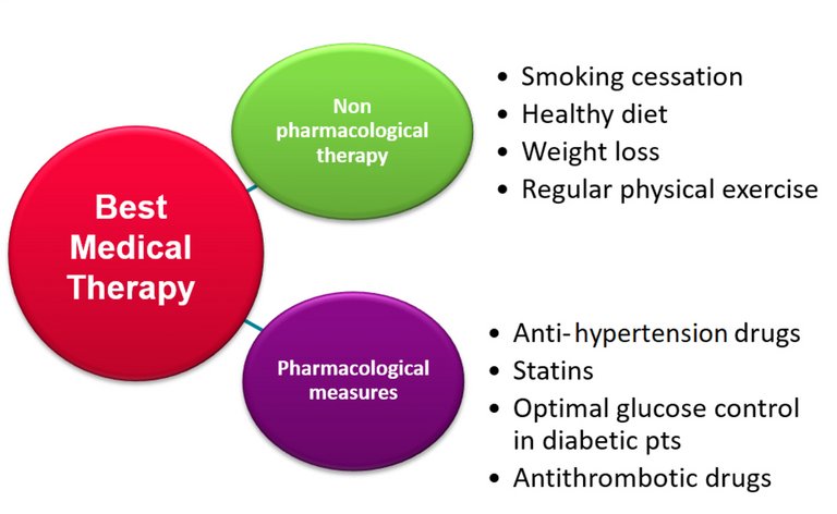
All patients with LEAD, regardless of their stage, should be managed with “best medical therapy” that includes management of cardiovascular risk factors, pharmacological therapy, and non-pharmacological measures such as smoking cessation, healthy diet, weight loss and regular physical exercise (fig. 1). However, in addition to best medical therapy, patients with incapacitating functional impairment or CLTI require urgent revascularisation to prevent amputation. In this subgroup, exercise training is not a first-line treatment.
Figure 1
Optimal medical treatment of lower extremity arterial disease.
Pharmacological measures
Pharmacological treatment includes antihypertensive, lipid-lowering and antithrombotic drugs, as well as optimal blood glucose control in diabetic patients.
In patients with LEAD, the use of statins has a level I-A recommendation, having been proved to improve maximum walking distance, reduce all-cause mortality and cardiovascular events, and limit adverse limb events [9].
According to the latest recommendations, a more aggressive reduction of low-density lipoprotein cholesterol across cardiovascular risk categories is warranted. Reduction of >50% from baseline levels and a goal of <1.8 mmol/l is recommended for high-risk, and <1.4 mmol/l for very high-risk patients [10].
For treatment of hypertension, angiotensin converting enzyme inhibitors (ACEIs), angiotensin receptor blockers (ARBs) or calcium antagonists should be preferred even in patients with CLTI, because of their potential for peripheral arterial dilatation (evidence IIa-B) [11]. Beta-blockers are not contraindicated in patients with LEAD, but they should be used carefully in CLTI patients [12].
Single antiplatelet therapy (SAPT) is not recommended for primary prevention in asymptomatic patients with only one arterial bed affected. Long-term SAPT is recommended in symptomatic patients and in those who have undergone revascularisation, and clopidogrel may be preferable to aspirin (evidence II-B). Dual antiplatelet therapy is recommended after infrainguinal stent implantation for at least 1 month and a longer duration in selected high-risk patients.
In patients with LEAD requiring long-term oral anticoagulation for other reasons, triple therapy, consisting of oral anticoagulation in addition to dual antiplatelet therapy, is not encouraged. For these patients, monotherapy with oral anticoagulation is indicated for asymptomatic LEAD or after open surgery (evidence IIa-B); additional SAPT (1 month) is recommended after revascularisation if its benefit outweighs the risk of bleeding complications. Prolonged SAPT in addition to a direct oral anticoagulant (DOAC) is indicated in high ischaemic risk patients. Triple therapy (DOAC + dual antiplatelet) should not be prescribed (evidence IIa-C) [13].
The Cardiovascular Outcomes for People Using Anticoagulation Strategies (COMPASS) trial, a multicentre randomised trial published in 2018, included patients with peripheral artery disease (PAD: previous revascularisation, amputation or symptomatic PAD), and found that low-dose rivaroxaban (2.5 mg twice a day) in combination with 100 mg aspirin significantly reduced MACEs (death, myocardial infarction or stroke) and major adverse limb events (MALEs) such as amputation and acute limb ischaemia, as compared with aspirin alone [14]. This drug combination was associated with a small but statistically significant increase in clinically relevant bleeding.
The sub-group of patients undergoing peripheral revascularisation was analysed in an international randomised, double-blind, placebo-controlled trial (VOYAGER PAD), which aimed to evaluate the efficacy and safety of this drug combination compared with 100 mg aspirin alone. Compared with aspirin alone, in these patients rivaroxaban + aspirin was associated with a significantly lower incidence of the composite outcome of acute limb ischaemia, major amputation for vascular causes, myocardial infarction, ischaemic stroke, or death from cardiovascular causes. The incidence of intracranial haemorrhage or fatal bleeding did not differ significantly between the groups [15].
Based on these results, the ESVM in its latest PAD guidelines recommend considering the combined treatment (rivaroxaban 2.5 mg twice daily + aspirin 100 mg once daily) in PAD patients without a high risk of bleeding [16].
Although study results are promising, the benefits and risks of the combination of low-dose rivaroxaban with low-dose aspirin in patients with CLTI are yet to be adequately defined.
Non-pharmacological measures
An appropriate lifestyle and Mediterranean diet with limited salt intake (<5–6 g/day) are recommended (evidence I-C).
The benefits of smoking cessation in reducing cardiovascular events and mortality are widely proven, especially in patients with cerebrovascular disease and LEAD (evidence I-B). Passive smoking should also be assessed and prevented [17].
Of great importance is supervised exercise therapy, which has proven benefits in terms of effort toleration and increased maximum and pain-free walking distance in patients with intermitted claudication, even if the ABI does not increase. Improvement is observed up to 2 years after a 3-month supervised programme. It should be proposed to every patient with LEAD (evidence I-A). Moreover, exercise subjectively improved the quality of life. Whether exercise training reduces cardiovascular events and improves life expectancy is still unclear. If supervised training is not available, unsupervised exercise should nonetheless be organised, although it less effective, because it has proven to be better than walking advice alone (evidence I-C) [18].
Alternative exercise modalities, such as cycling, strength training and upper-arm ergometry have also been shown to be effective and represent an alternative when walking training is not an option for functional reasons. Supervised schemes are mostly based on a 3-month programme, with a minimum of 3 hours/week, and a schedule of various walking and other exercises. Improved balance, strength and quality of life are reported, in addition to a gain in maximum pain-free walking distance. Although more cost effective than unsupervised exercise training, it is not reimbursed or available in every country.
As stated before, exercise is of paramount importance in patients with intermittent claudication, but should not be proposed to CTLI patients as first-line treatment to avoid a delay in revascularisation.
Conclusions
Management of LEAD is evolving over time and a several studies are ongoing. LEAD still represents a major public health concern, as the prevalence of this disease is high and growing.
Control of cardiovascular risk factors is of paramount importance and is achieved more and more effectively with a combination of pharmacological and non-pharmacological measures, confirming optimal medical treatment as a cornerstone of LEAD management.
Key points
When a vascular territory is affected by atherosclerosis, not only is the corresponding organ endangered, but the total risk of any cardiovascular event is also increased.
Thorough clinical history and physical examination are key steps in LEAD management.
ABI measurement is indicated in all patients suspected of having LEAD because of pulse abolition, intermittent claudication, or a non-healing extremity wound, and in all patients diagnosed with coronary artery disease, any peripheral artery disease, aortic aneurysm, chronic kidney disease or heart failure. In addition to its diagnostic value in LEAD, ABI is a strong marker for cardiovascular events.
Best medical therapy includes cardiovascular risk factor management, optimal pharmacological therapy and as non-pharmacological measures such as smoking cessation, healthy diet, weight loss and regular physical exercise.
Single antiplatelet therapy is indicated for LEAD patients who are symptomatic, have atherosclerosis involving several locations, or have undergone revascularisation. Clopidogrel is the preferred antiplatelet drug in LEAD patients. Statins are recommended, with the strongest level of evidence, for all patients with LEAD.
Correspondence
Professor Lucia Mazzolai, MD, PhD
Centre Hospitalier Universitaire Vaudois
Service d'angiologie
Av. Pierre-Decker 5
CH-1011 Lausanne
lucia.mazzolai[at]chuv.ch
References
1Aboyans V, Ricco JB, Bartelink MEL, Björck M, Brodmann M, Cohnert T, et al.; ESC Scientific Document Group. 2017 ESC Guidelines on the Diagnosis and Treatment of Peripheral Arterial Diseases, in collaboration with the European Society for Vascular Surgery (ESVS): Document covering atherosclerotic disease of extracranial carotid and vertebral, mesenteric, renal, upper and lower extremity arteries. Endorsed by: the European Stroke Organization (ESO)The Task Force for the Diagnosis and Treatment of Peripheral Arterial Diseases of the European Society of Cardiology (ESC) and of the European Society for Vascular Surgery (ESVS). Eur Heart J. 2018;39(9):763–816. doi:. http://dx.doi.org/10.1093/eurheartj/ehx095PubMed
2Fowkes FG, Rudan D, Rudan I, Aboyans V, Denenberg JO, McDermott MM, et al. Comparison of global estimates of prevalence and risk factors for peripheral artery disease in 2000 and 2010: a systematic review and analysis. Lancet. 2013;382(9901):1329–40. doi:. http://dx.doi.org/10.1016/S0140-6736(13)61249-0PubMed
3Giannopoulos A, Kakkos S, Abbott A, Naylor AR, Richards T, Mikhailidis DP, et al. Long-term mortality in patients with asymptomatic carotid stenosis: implications for statin therapy. Eur J Vasc Endovasc Surg. 2015;50(5):573–82. doi:. http://dx.doi.org/10.1016/j.ejvs.2015.06.115PubMed
4Abu Dabrh AM, Steffen MW, Undavalli C, Asi N, Wang Z, Elamin MB, et al. The natural history of untreated severe or critical limb ischemia. J Vasc Surg. 2015;62(6):1642–51.e3. doi:. http://dx.doi.org/10.1016/j.jvs.2015.07.065PubMed
5McKenna M, Wolfson S, Kuller L. The ratio of ankle and arm arterial pressure as an independent predictor of mortality. Atherosclerosis. 1991;87(2-3):119–28. doi:. http://dx.doi.org/10.1016/0021-9150(91)90014-TPubMed
6Fowkes FG, Murray GD, Butcher I, Heald CL, Lee RJ, Chambless LE, et al., Ankle Brachial Index Collaboration. Ankle brachial index combined with Framingham Risk Score to predict cardiovascular events and mortality: a meta-analysis. JAMA. 2008;300(2):197–208. doi:. http://dx.doi.org/10.1001/jama.300.2.197PubMed
7Belcaro G, Nicolaides AN, Ramaswami G, Cesarone MR, De Sanctis M, Incandela L, et al. Carotid and femoral ultrasound morphology screening and cardiovascular events in low risk subjects: a 10-year follow-up study (the CAFES-CAVE study(1)). Atherosclerosis. 2001;156(2):379–87. doi:. http://dx.doi.org/10.1016/S0021-9150(00)00665-1PubMed
8Aboyans V, Ricco JB, Bartelink MEL, Bjorck M, Brodmann M, Cohnert T, et al. 2017 ESC Guidelines on the Diagnosis and Treatment of Peripheral Arterial Diseases, in collaboration with the European Society for Vascular Surgery (ESVS). Rev Esp Cardiol (Engl Ed). 2018;71(2):111. doi:. http://dx.doi.org/10.1016/j.rec.2017.12.014PubMed
9Westin GG, Armstrong EJ, Bang H, Yeo KK, Anderson D, Dawson DL, et al. Association between statin medications and mortality, major adverse cardiovascular event, and amputation-free survival in patients with critical limb ischemia. J Am Coll Cardiol. 2014;63(7):682–90. doi:. http://dx.doi.org/10.1016/j.jacc.2013.09.073PubMed
10Mach F, Baigent C, Catapano AL, Koskinas KC, Casula M, Badimon L, et al.; ESC Scientific Document Group. 2019 ESC/EAS Guidelines for the management of dyslipidaemias: lipid modification to reduce cardiovascular risk: The Task Force for the management of dyslipidaemias of the European Society of Cardiology (ESC) and European Atherosclerosis Society (EAS). Eur Heart J. 2020;41(1):111–88. doi:. http://dx.doi.org/10.1093/eurheartj/ehz455PubMed
11Shahin Y, Barnes R, Barakat H, Chetter IC. Meta-analysis of angiotensin converting enzyme inhibitors effect on walking ability and ankle brachial pressure index in patients with intermittent claudication. Atherosclerosis. 2013;231(2):283–90. doi:. http://dx.doi.org/10.1016/j.atherosclerosis.2013.09.037PubMed
12Paravastu SC, Mendonca DA, Da Silva A. Beta blockers for peripheral arterial disease. Cochrane Database Syst Rev. 2013;(9):CD005508. PubMed
13CAPRIE Steering Committee. A randomised, blinded, trial of clopidogrel versus aspirin in patients at risk of ischaemic events (CAPRIE). Lancet. 1996;348(9038):1329–39. doi:. http://dx.doi.org/10.1016/S0140-6736(96)09457-3PubMed
14Anand SS, Caron F, Eikelboom JW, Bosch J, Dyal L, Aboyans V, et al. Major adverse limb events and mortality in patients with peripheral artery disease: the COMPASS trial. J Am Coll Cardiol. 2018;71(20):2306–15. doi:. http://dx.doi.org/10.1016/j.jacc.2018.03.008PubMed
15Bonaca MP, Bauersachs RM, Anand SS, Debus ES, Nehler MR, Patel MR, et al. Rivaroxaban in Peripheral Artery Disease after Revascularization. N Engl J Med. 2020;382(21):1994–2004. doi:. http://dx.doi.org/10.1056/NEJMoa2000052PubMed
16Frank U, Nikol S, Belch J, Boc V, Brodmann M, Carpentier PH, et al. ESVM Guideline on peripheral arterial disease. Vasa. 2019;48(Suppl 102):1–79. doi:. http://dx.doi.org/10.1024/0301-1526/a000834PubMed
17Joosten MM, Pai JK, Bertoia ML, Rimm EB, Spiegelman D, Mittleman MA, et al. Associations between conventional cardiovascular risk factors and risk of peripheral artery disease in men. JAMA. 2012;308(16):1660–7. doi:. http://dx.doi.org/10.1001/jama.2012.13415PubMed
18Bäck M, Jivegård L, Johansson A, Nordanstig J, Svanberg T, Adania UW, et al. Home-based supervised exercise versus hospital-based supervised exercise or unsupervised walk advice as treatment for intermittent claudication: a systematic review. J Rehabil Med. 2015;47(9):801–8. doi:. http://dx.doi.org/10.2340/16501977-2012PubMed